Metaplasia of the cervix
Metaplasia of the cervix is \u200b\u200bone of the precancerous diseases that has serious consequences, in the absence of treatment or incorrect diagnostic and therapeutic tactics. This pathology is common in women over 50, although it happens earlier. But not all women have a clue about what in question, if this diagnosis is established. Due to the fact that metaplasia of the cervix with late diagnosis tends to one hundred percent malignancy in the future, it is better to prevent this pathology at the stage of initial changes. For this purpose, all women at the stage of the usual daily routine examination by a gynecologist undergo a screening examination for metaplasia.
Causes of cervical metaplasia
Speaking about the development of cervical dysplasia, it is very important to highlight the main risk factors and etiological reasons for the development of this process.
Among the etiological factors, that is probable causes development of pathology, in the first place are infectious agents. Potential pathogens include both viruses and bacteria. Among viral agents, this is most often a woman's infection with the human papillomavirus. This virus has a tropism for the female genital organs and causes the development of another disease - condyloma or papilloma of the cervix. But infection long time may not make itself felt, and its course may be asymptomatic, and in some cases, cervical dysplasia may develop. Other possible agents are herpes viruses of all types. These viruses also have a tropism for the epithelium of the cervix and a sufficiently high oncogenicity, so they can be a trigger for the development of dysplastic processes in the cell.
Bacteria are less likely to play a role in the development of this disease, since they do not penetrate the cell's nuclear apparatus and do not induce changes in the genetic material. But among the possible bacterial infections only intracellular ones are of greater importance - these are ureoplasma, toxoplasma, chlamydia, gonococcus. These microorganisms penetrate into the cell and remain there for a very long time, while defending themselves against immune cells and maintaining a chronic focus of inflammation. This is not true reason dysplasia, but against its background, similar changes can develop, which will lead to dysplasia in the future.
The causes of cervical metaplasia are difficult to establish precisely, but today, one of the proven etiological factors is infection with the human papillomavirus, which plays a key role in the further progression of changes inside the cell.
Risk factors
Risk factors can be categorized into general and local. Common include bad habits, smoking, alcohol consumption, eating disorders and consumption of carcinogenic products, influence of factors environment... All these changes are accompanied, first of all, by a decrease in the reactivity of the whole organism, and against this background, functional and then morphological changes of organs and systems develop.
Among the risk factors for the development of cervical dysplasia are local - early start sex life, frequent change sexual partners, as well as infectious and inflammatory diseases of the female genital organs, frequent surgical interventions - abortion, disorders hormonal background, traumatic injuries.
Pathogenesis
Speaking about the development of dysplastic processes, it is very difficult to identify the period when they develop and the duration of the course of such changes. Since pathology can also occur in women under 40, it is important to be vigilant if metaplasia is suspected.
The structure of the cervix in healthy woman Is the alternation of the epithelial cover:
- squamous stratified non-keratinizing epithelium - located in the endocervix closer to the vaginal canal and is a continuation of it;
- intermediate zone - located further and is the border on the way to the cervix, both types of epithelium are absent here;
- columnar epithelium - lines the cervical cavity and cervical canal.
Normally, these balls do not mix and are clear border between them.
The pathogenesis of the development of cervical metaplasia begins with a triggering factor, which may be a viral agent. In this case, the virus penetrates into the cell, where its nucleic acid enters the nucleus by disrupting the integrity of the nuclear envelope.

As a result, the genetic apparatus of a normal cell is disrupted and the virus initiates the synthesis of its own proteins, which are necessary for it to function. It disrupts normal life cycle epithelial cell and its processes of division and reproduction. This is how abnormal cell division is formed, which contributes to the appearance of epithelial cells with nuclear atypia. That is, the process of cell division can stop at a certain phase of mitosis, and then the development of numerical cells with an incorrect set of chromosomes can be initiated. Such cells cannot provide normal protein synthesis and metabolism in the cytoplasm, which is the reason for the appearance of dysplastic processes in the cell. Such cells multiply and can move from their main location - for example, the cylindrical epithelium extends beyond the intermediate zone and zones appear among the normal squamous epithelium of the endocervix columnar epithelium, which is the phenomenon of metaplasia. Such a violation of the normal structure of the epithelial cover does not reach the basement membrane.
Today the definition of metaplasia or dysplasia is outdated, and a new term is used - CIN - cervical intraepithelial neoplasia. This concept makes it possible to clarify that this process is not so much dysplastic as pre-cancerous.
Symptoms of cervical metaplasia
Given that this pathology often passes without symptoms, it is necessary to know the main symptoms that allow you to pay attention and suspect this problem.
Metaplasia of the cervix is \u200b\u200ba dangerous condition precisely for the reason that often the morphological changes that develop are much ahead of the development of at least minimal clinical symptoms. This is one of the reasons for the introduction mandatory screening this pathology.
Symptoms of cervical metaplasia can occur most often with some provoking factor. This can be with concomitant warts, erosions, infectious lesions. Less often clinical picture occurs without such conditions and is characterized by pain during intercourse, a violation of the normal menstrual cycleif there are hormonal disruptions, vaginal discharge. Discharge can be in the form of leucorrhoea - curdled, copious, white or milky discharge with unpleasant odor, as well as in the form of bloody discharge before menstruation, after it or after intercourse. Local pain in metaplasia is not typical if it is a purely dysplastic process.
The first signs that appear most often and are not specific, but should be alerted, are painful intercourse. Unpleasant sensations arise as a result of injury to the dysplastic epithelium, which can also be accompanied by bloody discharge... This does not happen very often, but it can be one of the first manifestations. As for older women, their first symptoms of metaplasia may often not be expressed due to involutive processes in the uterus and cervix, which suggests premenopausal changes. So the symptoms that appear in a woman, she explains by the onset of menopause and does not go to the doctor.
Given that the clinical symptoms are not sufficiently pronounced, it is necessary to take any changes in the state of health very seriously, especially in older women.
Forms
Since the epithelium of the cervix has several types of cells, metaplasia can also be different. The histological picture of smear changes is studied to make the correct diagnosis and choose treatment tactics. In this case, not only the degree of distribution of atypical cells is indicated, but also the nature of these changes and the morphological features of the smear.
There are several types of dysplasia:
- immature metaplasia of the cervix;
- squamous metaplasia of the cervix with dyskaryosis;
- squamous metaplasia of the cervix.
Concerning immature metaplasia, then this is considered the most unfavorable option, since the more low level cell differentiation, the more the risk of malignancy increases. The picture of immature dysplasia is characterized by the appearance in the smear of cells that are small in size, indistinct unequal boundaries, and are also randomly located in the smear itself. Concerning internal structure cells, then the cytoplasm is changed with a violation of the location and structure of the structural elements of the cell. Various changes in mitoses are observed in the nucleus. Sometimes it is difficult to attribute such cells to any type of epithelium, since they do not have characteristic distinctive features.
Squamous metaplasia cervical with dyskaryosis is a more differentiated species compared to immature forms. Such cells already have a certain shape, the same size and a sufficient amount. Inside the cell, the cytoplasm is not changed, and the structural elements are located correctly, in sufficient quantities, which characterizes the cytoplasm of a normal epithelial cell. The only differences from normal cells are abnormal divisions in the nucleus in the form of pathological mitoses. This is what characterizes the term "dyskaryosis".
Squamous metaplasia of the cervix - This is the most differentiated option, since the epithelium has all the characteristics of normal cells, with the exception of location. So with squamous metaplasia of the cervix, the squamous stratified epithelium is determined behind the intermediate zone in the region of the cervical canal among the columnar epithelium.
These histological types do not affect the course of the disease, but have a different prognosis, therefore such a classification is mandatory in cytological research.
Diagnosis of cervical metaplasia
Since the course of this pathology is often asymptomatic, then important element timely diagnosis and prevention of complications are preventive examinations by a gynecologist, which a woman must undergo annually. During the examination, the doctor examines cervix women in mirrors, which makes it possible to see changes that can be seen without complementary methods... Several cells of the metaplastic epithelium among the normal cover, as a rule, are not visible, therefore, a mandatory stage of the examination is taking a smear with a special brush for histological examination and detection of dysplasia.
Must be observed the right technology - a smear is taken from three zones of the cervix - the endocervix, the intermediate zone and the cervical canal, that is, all three types of epithelium must be present. This is where the objective examination ends. Further, all smears are sent to the laboratory for cytology and histology.
The tests that the doctor receives from the laboratory suggest dysplastic changes. There are six main types of strokes:
- histological picture of a healthy woman;
- inflammatory and benign changes in the smear;
- cervical intraepithelial neoplasia
- light metaplasia (CIN-I) - altered dysplastic cells extend into the depth of no more than one third of the epithelial cover;
- moderate metaplasia (CIN-II) - altered dysplastic cells extend into the depth by no more than two-thirds;
- severe metaplasia (CIN-ІІІ) altered dysplastic cells spread deeper by two-thirds or more, but without invasion of the basement membrane;
- suspected cancer;
- uninformative smear (not all types of epithelium are presented).
If a smear comes and the woman is healthy, everything is fine, but in all other cases, the woman is called for a second consultation and further examination.
If metaplasia is suspected, that is, if the smear is of the third type, then instrumental research methods are performed.
The epithelium lining the upper part of the female genital tract (tubal, endometrial, and endocervical) develops from the Müllerian (parameso-nephral) duct. Despite the fact that each organ has its own epithelium, the epithelium of the Müllerian duct can be found everywhere. Thus, although the typical endocervical epithelium is predominantly built of tall, cylindrical mucin-secreting cells with basal nuclei, there are glands or groups of glands lined with tubal or endometrial epithelium. Also, endometrial (endometrial-like) cells or mucinous epithelium can be found in the fallopian tube. These variants of the Müllerian epithelium should not be considered as a disease or as metaplasia, since this is just an incorrect differentiation of the Müllerian duct epithelium.
The presence of tubal epithelium in the cervix is \u200b\u200bcalled tubal metaplasia (endosalpingosis), endometrial type - endometrioid metaplasia; there is also a mixed variant - tuboendometrioid metaplasia. The described changes are found in 69% of cases of conization, 70% of hysterectomies for benign processes and 89% of hysterectomies for squamous cell carcinoma of the cervix. It was revealed that the normal uterine glands of the tuboendometrial type continue from the lower segment of the uterus towards the vaginal portion of the cervix, forming a sleeve located deeper than the cervical mucinous glands. The version is also discussed that normal neck The uterus contains two layers of mesenchyme with its own separate epithelium. The superficial layer supports mucinous differentiation of the epithelium, and the second (deeper) layer, which serves as a continuation of the mesenchyme of the body of the uterus and vagina, contains submerged glands from the tuboendometrial epithelium, which clutch-like cover the cervix, continuing from the endometrium. It is important to know about the normal presence of tuboendometrial glands, since they can be mistakenly regarded as dysplasia of the glandular epithelium.
Of all the variants of Müller's "metaplasia", the most common is tubal metaplasia (endosalpingosis). It is characterized by the presence of structurally normal glands in the stroma of the cervix, lined with cells resembling the epithelium of the fallopian tube. All types of cells are found: light (ciliated) cells, cells without cilia, and intercalary cells. Tubal metaplasia is usually a single gland or a group of glands. Slightly less common is the mixed variant - tuboendometrioid metaplasia. And extremely rarely there is a "pure" endometrioid metaplasia, represented by single or multiple glands. Müllerian "metaplasias" are usually asymptomatic and are an accidental finding in hysterectomies performed for other indications. However, when superficially located, metaplastic cells can get into the smear and, therefore, be treated as atypical.
A pseudo-infiltrative type of tubal metaplasia has been described. Due to the scattered arrangement of the glands given view metaplasia must be differentiated from malignant adenoma. For pseudo-infiltrative tubal metaplasia, nuclear atypia and desmoplastic stromal reaction are uncharacteristic. In three cases described, the use of diethylstilbestrol took place. It is possible that the pseudo-infiltrative nature of tubal metaplasia is a form of diethylstilbestrol-associated adenosis in the cervix.
Intestinal metaplasia
A rare form of metaplasia that occurs in the cervix and is characterized by the appearance of single goblet cells that replace the normal mucin-producing epithelium of the cervical canal, and crypts.
Along with the goblet cells, argentaffin cells are also found in the epithelium. Intestinal metaplasia is often combined with dysplasia of the glandular epithelium. Sometimes goblet cells squeeze and deform the nuclei of adjacent cells, which makes diagnosis difficult. Intestinal metaplasia also occurs in foci of adenocarcinoma in situ (intestinal type AIS).
Atypical oxyphilic metaplasia
Most often it is an accidental finding on microscopy and has no clinical significance. Changes in the glands are local in nature, similar to apocrine metaplasia. The lining is represented by cubic cells, lying in one layer, with a brightly oxyphilic extensive cytoplasm and an apical protrusion; nuclei can be hyperchromic, uneven, segmented. Stratification, proliferative activity and atypia are not detected. Often, atypical oxyphilic metaplasia is combined with inflammatory changes.
Squamous (squamous) metaplasia is a term used to describe cellular noncancerous changes in the epithelial lining of certain internal organssuch as the bladder, cervix and lungs. Metaplasia occurs when constant stress or irritation causes a reversible process in which differentiated epithelial cells of one type are converted to epithelial cells of another type. In squamous metaplasia, various epithelial cells are replaced as a result of the adaptation mechanism by the squamous epithelium.
A change in cell type can lead to a decrease in epithelial function. When abnormal stimuli are removed, metaplastic cells return to their original form and function. The persistence of physiological stressors in areas prone to metaplasia can lead to dysplasia or.
Dysplastic cells, in the absence of timely elimination of the stressor or irritant, can transform into cancer cells.
The epithelium is composed of layers of various epithelial cells such as cylindrical, cubic, and flat cells. Cylindrical epithelial cells are elongated, cylinder-shaped cells that form the lining of the cervical canal (endocervix), intestine, and stomach. Cylindrical cells with nuclei at different heights are called pseudostratified epithelial cells. Cubic epithelium is formed from square-shaped cells. Such cells are commonly found in exocrine glands and renal tubules. Squamous epithelial cells, in turn, form smooth epithelium.
The process of development of squamous cell metaplasia and the resulting neoplastic changes in cells can be well seen on the example of cervical metaplasia. The mucous membrane of the cervical canal, which is a cavity in the cervix, is usually formed by columnar epithelial cells. Estrogen and constant exposure to acidic pH levels in the vagina provoke a process of squamous metaplasia that affects the epithelium of the cervical canal. In response to irritating pH levels, fragile columnar epithelial cells begin to be replaced by stronger flat cells.
Squamous metaplasia of the cervical canal also occurs as a result of fusion with the adjacent ectocervix.
Squamous cells of the ectocervix begin to fill the endocervical area, replacing columnar epithelial cells. When carcinogenic factors such as the presence of human papillomavirus act as stimuli or stressors on metaplastic epithelial cells, a cancer of the cervix is \u200b\u200bformed. A similar metaplastic process occurs when cigarette smoke constantly irritates the pseudostratified epithelial cells of the lung mucosa.
Cigarette smoke is a stressor that transforms pseudo-stratified cells into much more hardy flat cells. However, even flat cells, for example, in the bladder, can undergo squamous metaplastic changes. Triangle bladder, or its inner triangular region, includes the squamous epithelial cells that form the lining of this region. When adult flat bladder cells undergo chronic inflammation, metaplastic changes in these cells occur.
Content
Metaplasia of the cervix often leads to the appearance of cancerous growths. In case of untimely diagnosis and absence drug therapy possible negative consequences... The presented pathology in most cases develops in women over 45 years old. But sometimes patients also turn to doctors at a young age.
With the progression of cervical metaplasia, there is a possibility of malignancy in the future, so it is important to determine the disease at the initial stage. That is why it is necessary to regularly visit your doctor for examination.
Causes of cervical metaplasia
The main factor in progression metaplasia of the cervix is \u200b\u200ba pathogenic bacteria.
In most cases, patients are diagnosed with the papilloma virus, which can be traced in the female genital organs. As a result of this effect, papillomas and condylomas of the cervix develop.
Long-term infection can affect the cervix without clear signs. After the penetration of bacteria into the body, they can cause the development of ureoplasma, gonococci and chlamydia. The latter penetrate into cells and remain in them for a long time.
The reasons for the development of cervical metaplasia are difficult to determine, but scientists have proved that main factor - This is the defeat of the human papillomavirus. It plays an important role in the progression of changes in cell tissues.

Risk factors
Are common
These include bad habits, alcoholic beverages, unhealthy diet, poor environmental situation and the use of carcinogenic products. During pathological changes in the body, activity and protective reaction decrease. Morphological and functional changes occur inside the body.
Local
This group of risk factors includes early onset of sexual activity, promiscuous sex life, infectious and inflammatory diseases of the female genital organs, frequent mechanical abortions, disruption of the normal hormonal background and traumatic injuries.
Pathogenesis of cervical metaplasia
Pathological changes are observed in patients different ages, therefore, when the first signs appear, it is important to immediately seek help from a doctor. A healthy epithelial lining of the cervix has the following structure:
- squamous stratified non-keratinizing epithelium - it is located near the vaginal canal and is its continuation;
- intermediate zone - this layer is located near the cervix;
- columnar epithelium - it lines the entire cavity of the cervix and cervical canal.
If pathogenic bacteria do not infect the cervix, then all layers do not mix with each other, and a clear boundary is clearly visible.
After the penetration of the viral agent, a disease such as cervical metaplasia begins to develop. Disease-causing bacteria penetrate into cells and the integrity of the nuclear membranes is disrupted. They begin chaotic division and epithelial cells appear with atypia of the nucleus. It is for this reason that it is important to start treatment on time.

Affected cells will not be able to provide normal protein synthesis inside the body, therefore dysplastic processes develop. Due to such violations, metaplasia of the cervix appears.
Symptoms
In most cases, metaplasia in the cervix occurs without obvious signs and symptoms. But every woman should know the common clinical manifestationsto detect in time pathological changes in the body and start treatment.
Cervical metaplasia means dangerous condition and the presence of morphological changes in the body.
For this reason, it is important for women to be screened regularly. With the progression of metaplasia of the cervix, intercourse becomes painful, and discomfort appears. This is due to the fact that the dysplastic epithelium begins to injure and bleed. During menopause, symptoms are less pronounced and women often confuse such a disease with menopause, so they do not seek help from a doctor.
Metaplasia causes genital warts, infectious diseases and cervical erosion. In women, discharge begins to increase, having a milky shade and a curdled consistency. In this case, a specific and characteristic smell appears.

Forms of metaplasia of the cervix
There are several forms of cervical metaplasia:
- immature;
- squamous;
- squamous cell metaplasia in combination with dyskaryosis.
To make a correct diagnosis, it is necessary to take vaginal swabs. During the diagnosis, specialists note the small size of cells and blurred boundaries. The cells of the cytoplasm are distinguished by a disturbed structure and the presence of structural elements.
Methods for diagnosing metaplasia of the cervix
Diagnose cervical metaplasia possible during a routine examination by a gynecologist.
A biopsy is required to confirm the diagnosis and identify malignant lesions. Doctors carefully examine all histological tissue types. If signs of malignant transformation are absent in the epithelial tissues, then doctors may not prescribe treatment.

Treatment is prescribed exclusively on an individual basis and after a comprehensive diagnosis. It is also important to take smears to assess the condition of the cells and their boundaries. Colposcopy may be required for additional examination. The doctor will study the structure of the cervix well using a special device that displays an image on the monitor screen. Thanks to this diagnostic method, the specialist will be able to examine in detail the areas of metaplasia that cannot be seen during a routine preventive examination.
Treatment methods
After confirming the diagnosis, the doctor will prescribe treatment to prevent the development of cancerous tumor.
Therapy can be conservative or operative, therefore, it is appointed exclusively on an individual basis.
Conservative treatment
Metaplasia of viral origin is treated medicineswhich have antiviral effects. The drugs suppress the activity of viruses and prevent them from multiplying.
If bacteria are present in the smear, it is advisable to prescribe treatment with antibacterial drugs. Patients take complex drugs - these are antibiotic and antifungal agents. Squamous metaplasia is treated with vaginal anti-inflammatory suppositories and medications that boost immunity.
Operative treatment
If drug treatment does not bring positive results, then the doctor decides on surgical intervention. It includes laser vaporization, cone exposure, electrocautery and curettage of the cervix. The choice of treatment depends on the testimony of the doctor and the results of the diagnosis received.

Metaplasia refers to serious diseases that develop in the cervix. This pathology requires treatment at the initial stage of development in order to prevent the appearance of a cancerous tumor. As a preventive measure, it is recommended to regularly visit a gynecologist for examination.
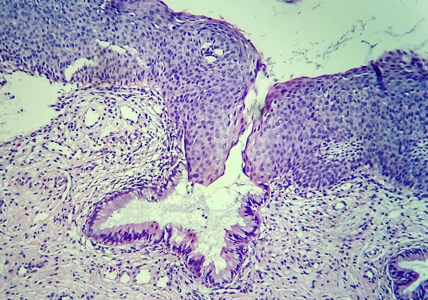
Squamous cell metaplasia of the cervix is \u200b\u200ban adaptive reaction of the body, characterized by the replacement of a more vulnerable single-layer columnar epithelium with a more resistant stratified squamous epithelium.
The junction of the cylindrical and stratified squamous epithelium
A single-layer cylindrical (aka glandular) epithelium lines the cervical canal of the cervix, trachea, bronchi and stomach. The inner lining of the uterus - the endometrium - also belongs to the glandular epithelium. This type of epithelium is quite delicate, located in one layer and is necessary for the secretion of mucus and other biologically active substances... Naturally, when exposed to glandular epithelium various irritating factors or when the hormonal background changes, the body is able to rebuild, adapt to new conditions by replacing the type of epithelium. The stratified squamous epithelium is several times thicker than the glandular epithelium and, accordingly, is more resistant to irritating factors. It covers the mucous membranes of the mouth, esophagus, vagina and the outer part of the cervix, as well as the skin. Squamous metaplasia is a process when squamous epithelium appears in unusual places or with functional displacement of the epithelium.
Squamous metaplasia of the cervix
As mentioned above, the cervical canal is lined with glandular epithelium, and the outer part of the cervix is \u200b\u200bflat. The epithelial junction (pictured above) can migrate at different distances within the cervix. This process is mainly controlled by hormones. So, in children, the epithelial junction is located near the external pharynx, while in older women it moves deep into the cervical canal. In women of reproductive age with various hormonal disorders, pregnancy, inflammatory diseases of the pelvic organs, the glandular epithelium can move beyond the external pharynx and even reach the fornix of the vagina. Gynecologists call this cervical erosion or cervical ectopia. With the normalization of the hormonal background, the glandular epithelium is again replaced by stratified squamous epithelium by squamous metaplasia (or epidermisation).
Epidermisation of the gland
Immature squamous metaplasia - the initial stage of metaplasia, characterized by the multiplication of germ (reserve) cells and their differentiation into squamous epithelium. Immature squamous epithelium contains little glycogen and is easily identified by cytological and histological examination. One of key points in diagnosis is the definition of cellular atypia, which is a sign of tumor transformation. Exclusion is the main task cytological examination... Reactive changes in the metaplastic epithelium are often associated with severe inflammatory changes in the smear.
Mature squamous metaplasia — the final stage metaplasia with the formation of a well-organized epithelial layer. Mature squamous epithelium contains a large number of glycogen and a lot of cytoplasm. This option is the most favorable.
Both the integumentary glandular epithelium and the epithelium of the glands undergo metaplasia. In this case, the excretory ducts of the glands can be blocked, which leads to the formation.
Squamous cell metaplasia does not require special treatment, it is enough just to exclude provoking factors and regularly undergo preventive examinations.
Squamous metaplasia in other organs
Squamous metaplasia is also observed in the lungs, namely in the bronchi of smokers, with chronic bronchitis or bronchiectasis. When long-term exposure carcinogenic factors, metaplastic squamous epithelium can undergo malignant transformation with the development of squamous lung cancer... Squamous metaplasia of the bronchi is an unfavorable process, therefore, to exclude provoking factors is the initial task in treatment.
Squamous metaplasia of the bladder - a frequent occurrence with chronic inflammation and looks like white spot against a pink background of normal mucosa. In this case, a person may experience various disorders in the work of the bladder, but the symptoms are nonspecific. Diagnostics is based on cytoscopy - examination of the bladder cavity using a special probe with a camera.
Squamous metaplasia of the epithelium can also be observed in polyps of the cervix and in adenocarcinoma. These are concomitant changes and without treatment of the underlying pathology, the effect cannot be achieved.