It is a pathological formation protruding above the mucous membrane. Such an education does not pose a direct threat to a woman's life, but, as a rule, prevents pregnancy. Doctors say that in the absence of qualified treatment for the pathology, the polyp after a while can be transformed into cancerous tumor. At the moment, there are several methods of influencing these formations, but hysteroscopy is the most suitable option for removing a polyp.
Polyp hysteroscopy: about the procedure
The procedure is modern method diagnostics of the uterus and targeted removal of pathological formations of the mucosa. Unlike previous treatments, polyp removal cervical canal and the uterine cavity during hysteroscopy does not cause complications.
The essence of the procedure is to introduce a hysteroscope into the uterus, which is a flexible tube with an optical device (camera). Thus, during hysteroscopy (polypectomy), the doctor can visually examine the uterine mucosa for inflammation and formations. When polyps are found, their targeted removal is carried out.
Preparation for hysteroscopy of the uterine polyp
Before hysteroscopy, the doctor must explain to the patient the essence of the upcoming procedure, as well as choose the type of anesthesia. It is necessary to tell the doctor:
- about allergies to any medications;
- about taking medications or herbal supplements;
- about blood clotting problems.
As a rule, hysteroscopy of the endometrial polyp is performed after the end of menstruation, but no later than on the tenth day of the cycle. It is believed that it is during this period that the maximum effectiveness of the procedure can be achieved.
Before hysteroscopy, namely, removal, the patient is advised to refuse to eat and drink for 4-6 hours. A week before the procedure, it is better not to take anti-inflammatory and blood-thinning drugs. The procedure itself takes from 10 to 45 minutes and is performed under local or general anesthesia.
Removal of a uterine polyp during hysteroscopy
As a rule, the procedure goes as follows:
- the patient is given anesthesia, the type of which is negotiated in advance;
- the doctor inserts a hysteroscope into the uterine cavity;
- the uterine cavity is filled with carbon dioxide or liquid for better visualization;
- if, as a result of the examination of the uterus, endometrial polyps were detected, their targeted removal is carried out with an instrument attached to the hysteroscope;
- after removal of the polyp, a second examination of the mucosa is carried out for the remains of formations;
- if the polyp or its base is not completely removed, a second removal is performed followed by an examination.
Recovery after hysteroscopy
As a rule, hysteroscopy is performed on an outpatient basis. Recovery after removal of a polyp using hysteroscopy depends on the type of anesthesia used, but most often the patient does not have complaints. Occasionally, a woman may feel pain in the lower abdomen, reminiscent of menstrual cramps. Bleeding usually ends 2-3 days after the procedure. 
In most cases, patients return to normal life within 1-2 days after the operation. In the first week, it is strictly forbidden to use any medications without the consent of your doctor.
- Why is early treatment important?
- How is the procedure carried out?
- Stages of preparation: testing and additional studies
- Preparing the patient for surgery
- Operation progress
- Recovery period after surgery
Polyps are a pathology - neoplasms on the mucous membrane. The polyp protrudes above the wall of a certain organ, connects to the wall directly with the base or leg. The surface of the formation is both smooth and folded. In its color, it is graded from light pink to light yellow. hysteroscopy is the most effective method.
Why is early treatment important?
Often polyps are found in the area of the uterus and its cervix. Treatment of these formations should be carried out in a timely manner. The sooner a woman thinks about removing the polyp, the sooner she will allow herself to get rid of such unpleasant manifestations of the polyp as spotting not during the period of menstruation, pain in the lower abdomen or pain during intercourse.
Even in cases where endometrial polyps do not manifest themselves in any way, it is important to keep them under control. An endometrial polyp can degenerate into malignant tumors and lead to cancer. Hysteroscopy solves the problem of this disease most effectively. It is not only a method of treatment, but also diagnostics. After the operation, the tissues of the outgrowth are sent to special study to prevent the development of cancer.
Back to index
How is the procedure carried out?
With the help of the hysteroscopy procedure, it is carried out in compliance with the rules of asepsis and antisepsis. The doctor inserts a special video camera into the uterine cavity through the genital opening. This camera allows you to take a closer look inner surface uterus, cervix. Other endoscopic instruments are introduced through the genital organ, making it possible to accurately and accurately remove the neoplasm. Reviews about this procedure among women who have undergone it are extremely positive.
In some sources, the name of this operation is hysteroresectoscopy. During it, a special surgical instrument is used, which allows performing precise operations in the uterine cavity. That is, hysteroresectoscopy and hysteroscopy are two names for the same procedure.
Back to index
Stages of preparation: testing and additional studies
A gynecologist examines with mirrors. Such an examination allows the doctor to get an impression of the state of the organs. reproductive system person. It is very important to conduct a bacteriological examination during the preparation for the removal of polyps. The procedure is done to make sure that the woman does not have infectious diseases, in which hysteroscopy may be contraindicated.
Contraindications:
- Any inflammatory processes genital organs;
- Profuse uterine bleeding;
- Pregnancy;
- Stenosis or cancer of the cervix;
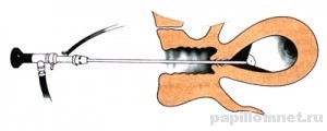
The image shows the approximate course of the operation.
If, after primary tests and examinations, the doctor prescribes an operation to remove it, then it is necessary to conduct an ultrasound scan of the pelvic organs. It is carried out through the genital organ. With the help of ultrasound, the doctor can determine the presence of polyps, their number and more accurate location. The results of the ultrasound will be important information for the specialist who will perform the operation.
Back to index
Preparing the patient for surgery
Preparation begins a few days before the operation. It is recommended to refrain from sexual intercourse. Douching should not be done for a week before surgery, use vaginal suppositories and sprays, tablets. All this is relevant if no other instructions have been received from the attending physician. The doctor prescribes the operation a couple of days after the end of menstruation.
Back to index
Operation progress
Requires hospitalization of the woman for several days. This procedure performed under local anesthesia. IN special occasions the use of general anesthesia is not ruled out.
A hysteroscope is a long, thin tube. At the end it has a video camera, a light source, a channel for inputting the appropriate instruments. The device is inserted through the cervix into the uterine cavity. This removes some air and fluid, which allows you to straighten the walls of the uterus and improve their examination.
The operation time is about half an hour. After the doctor finds the site of attachment of the polyp, he uses flexible scissors to cut off the legs of the outgrowth. Then the doctor fixes the removed polyp with forceps and carefully removes it from the uterine cavity. When endometrial polyps have big size, additional expansion of the channel with the help of Gegar expanders is required.
Polyps of the uterus are pathological neoplasms of the mucous membrane protruding above the wall of the uterus. Polyps are attached in the uterus with a base or stalk. Polyps can have a smooth or ribbed surface. The color of polyps may change from pale pink to light yellow. Often there are polyps with a pronounced vascular pattern.
The presence of polyps in the uterus, gives women a lot of problems and is characterized by the appearance spotting between menstrual cycles, pain in the lower abdomen at rest and during intercourse. For the treatment of this disease, it is important to conduct timely diagnosis and treatment.
Hysteroscopy as a method of removing polyps
Today, polyps are removed by hysteroscopy, which shows high efficiency. During the operation to remove polyps, simultaneous diagnosis is possible. Using an optical device, the doctor assesses the condition of the inner walls of the uterus, and the removed polyp can be sent for histological examination to confirm or exclude the possibility of cancer.
Hysteroscopy of the uterus
The hysteroscopy procedure is performed in compliance with the rules of asepsis and antisepsis. A hysteroscope is a medical optical device with a microvideo camera that a doctor inserts into the uterus through the vagina. This device allows the doctor to make an examination and assess the condition of the uterine cavity, and also makes the removal of the polyp extremely accurate.
Preliminary preparation for the hysteroscopy procedure
A gynecological examination of the patient with the help of mirrors and a manual examination of the internal genital organs are carried out before histology. In preparation for the hysteroscopy, a smear from the vagina must be examined for flora. This procedure is necessary in order to find out if the patient has infectious diseases, the presence of which will make hysteroscopy unsafe. Infectious diseases are a serious contraindication to the procedure.
If the doctor plans to remove the polyp from the uterine cavity, the patient passes ultrasound procedure pelvic organs. Conducting a transvaginal ultrasound makes it possible to determine whether there are polyps in the uterus, how many there are, what their size is, how they are located and attached to the walls of the uterus. Such information is very important for the doctor who will perform the hysteroscopy of the uterine polyp.
Also, an ultrasound scan in combination with a special test will make it possible to confirm or exclude the pregnancy of the patient who is prescribed hysteroscopy, polyp removal.
How to prepare for the operation?
During the week before the operation, the patient should not carry out douching procedures, use vaginal suppositories, exclude medicines(except as prescribed by the attending physician). It is necessary to exclude sexual intercourse a few days before the operation. The operation is prescribed for patients a few days after the menstruation has passed.
Polyp removal
To prepare and carry out an operation to remove a uterine polyp, the patient is placed in a hospital for several days. Hysteroscopy can be performed under local or general anesthesia, it all depends on the characteristics of the patient's body and the complexity of the disease.
A hysteroscope is a medical optical device consisting of a long probe equipped with a miniature video camera at the end. Also on the device there is a light source and a channel that allows the introduction of tools.
The introduction of the device is made through the cervix into the uterine cavity, which is then filled with air or fluid to straighten the walls of the uterus, so that it is easier to examine the uterine cavity.
The operation takes 30-40 minutes. When the polyp is found, the doctor cuts off the polyp leg with flexible hysteroscope scissors. The cut polyp is clamped with forceps and evacuated from the uterine cavity. If the polyps are quite large, in order to make it convenient to remove them from the cavity, the cervical canal is expanded with special Hegar dilators.
Recovery period
After the operation hysteroscopy - polyp removal will be completed, saved bleeding meager nature. There is also the appearance of moderate pain in the lower abdomen, discomfort and pain in the perineum. This should not be surprising, any surgical intervention, even if it refers to minimally invasive methods, is always a trauma. In the normal state of the body, the symptoms will disappear without medical intervention in a few days.
But there may be another picture of the postoperative period of hysteroscopy. The patient may experience heavy bleeding, uncharacteristic vaginal discharge, fever, severe pain. Inflammation or infection may occur. You do not need to make any attempts on your own, you should immediately seek help from a doctor in order to avoid undesirable consequences.
Restrictions after surgery
After the operation, you need to perform for some time necessary rules and requirements to reduce the risk of possible complications. For some time, you need to give up sexual intercourse, exclude the use hygienic tampons, do not take hot baths, do not visit the bath and sauna, so as not to provoke bleeding. Not allowed in postoperative period douche.
Hysteroscopy does not reduce a woman's fertility, as practice shows, pregnancy can occur in the next six months. Timely diagnosis and removal of polyps gives a woman the opportunity to fully bear a child, because often it is the presence