Pathology of the cervix is one of the most urgent problems and a frequent reason for patients to visit a gynecologist. Many women have heard that there are changes on the cervix: erosion, dysplasia, and even cancer. What preventive measures can be taken to prevent the occurrence serious problems? How are they diagnosed? Is it necessary to treat pathological conditions of the cervix, and what methods are most effective today?
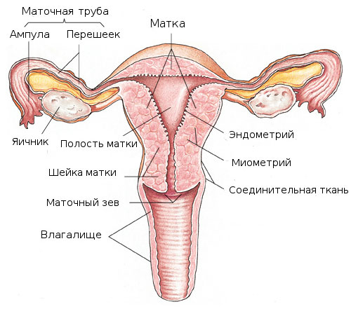
The cervix is \u200b\u200bone of the few organs of the female reproductive system available for visual inspection. This means that the occurrence oncological diseases the cervix can be completely prevented. Nevertheless, cervical cancer ranks first in the structure of oncological diseases of the female reproductive system.
Causes of diseases of the cervix:
- Injury (after abortion or childbirth).
- Infection (chlamydia, cocci, Trichomonas, mycolasma, etc.) cause inflammation and increased desquamation of the epithelium, under which there are immature cells with increased susceptibility.
- Viruses... Currently, a link has been proven between the occurrence of cervical cancer and the human papillomavirus. It should be noted that HPV exerts its oncogenic effect only in those tissues that are prepared by other infectious agents and the herpes simplex virus type 2.
- Hormonal imbalance (most often progesterone deficiency state).
- Violation of local immunity, which plays an important role in the processes of tissue repair and the occurrence of cervical erosion.
All diseases of the cervix can be divided into: background, precancerous (dysplasia) and cervical cancer.
Background processes: pseudo-erosion (ectopia, exactly what is most often called cervical erosion), leukoplakia without atypia, polyps, condylomas.
Background processes are not in themselves precancerous conditions, but pathological changes can develop against their background.
The causes of background processes are manifold - dyshormonal, inflammatory, post-traumatic. Very often, background processes overlap and create pictures that are difficult to diagnose.
Precancerous process (dysplasia): mild, moderate, severe.
Precancerous processes (dysplasia) are complexes of atypical cells that do not go beyond the border of the epithelial cover. Depending on the severity, mild, moderate and severe dysplasias are distinguished. The presence of dysplasia can be suspected during colposcopy and detected when cytological examination... Dysplasia is a mandatory step in the transition to cervical cancer. That is, cervical cancer rarely occurs on the unchanged cervix, it is usually preceded by dysplasia.
Separately distinguish inflammatory diseases cervix, but this is more related to genital infections.
The most common diseases of the cervix
- Cervical erosion in its pure form, it is characterized by damage to the mucous membrane of the cervix, facing the vagina. That is, this is a kind of wound on the cervix, visible to the naked eye when examined by a gynecologist. It looks like a bright red spot on a pink background of the intact mucous membrane of the cervix. Most often, the cause of the formation of erosion is the inflammatory process, various chemical or physical influences or hormonal disorders. A similar picture, a gynecologist can rarely observe, since this erosion usually exists for no more than two weeks. Later, if the cause is eliminated, the erosion heals completely. If healing does not occur, erosion turns into pseudo-erosion. It is precisely it that is most often designated by the term "cervical erosion".
- Pseudo-erosion, in turn, represents an incomplete or incompletely healed erosion of the cervix. That is, the healing began, but since the causative factor was not eliminated, the process went wrong. As a result of such "incorrect" healing, in particular, on the cervix, so-called nabot cysts are formed, which are also quite common.
- Nabotov cysts are clogged ducts of the glands of the cervix, stretched by the secretion of these glands. In simple terms, these are fluid-filled bubbles located under the mucous membrane on the surface of the cervix. Sometimes, if the cause of the erosion is eliminated, a complete cure can occur. But more often the process of erosion healing proceeds in waves, with a change in the picture visible upon examination. With a prolonged course of cervical erosion, the processes that constantly occur in the cells of the surface layer of the cervix can change, leading to the development of dysplasia. If this does not happen, the erosion of the cervix can exist for an arbitrarily long time, clinically not manifesting itself in any way.
- Leukoplakia can develop from erosion and is a seal on the cervix in the form white spot... Leukoplakia can also progress with the development of precancerous conditions.
- Polyps of the cervix - these are outgrowths on the surface of the neck or in its channel of various structures. The reason for the development of polyps is most often hormonal or inflammatory processes.
- Flat warts represent one of the manifestations of human papillomavirus infection. Dysplasia can also develop within flat warts, which characterizes them as precancer.
- Dysplasia of the cervix there are three degrees. The first is mild, the second is moderate and the third is severe dysplasia. The degrees differ depending on the depth of the lesion. The deeper the process, the more severe the dysplasia. Dysplasia is not visible to the naked eye and can only be determined when special studies both on the pathologically altered cervix and in healthy women.
The risk group for the development of various processes on the cervix can be attributed primarily to women who start early sex life, who often change sexual partners, who have given birth many times or have repeatedly undergone various intrauterine interventions.
Diagnostic tests
Oncocytological smear or PAP test is a very simple, painless, and at the same time informative diagnostic method malignant processes epithelium of the cervix. It is recommended to do it once a year.
Colposcopy - another way to assess the state of the cervix, which involves examining the structure of the epithelium under a microscope (colposcope). During the examination, the cervix is \u200b\u200bprocessed with special dye solutions (solution is used as reagents acetic acid, Lugol's solution and color filters). Thus, it becomes possible to identify pathology, if any, and determine its boundaries. The procedure is absolutely painless, since the magnification of the colposcope allows you to assess the condition of the cervix at a distance.
Cervical biopsy - the most informative and serious method for diagnosing the state of the epithelium. If, as a result of colposcopy, the doctor sees an area of \u200b\u200bthe epithelium that is suspicious of dysplasia or cervical cancer, then, in agreement with the patient, a piece of cervical tissue is taken for histological examination.
Treatment of diseases of the cervix
As for the treatment, then modern medicine offers a wide range of treatments for cervical diseases. First of all, it is necessary to establish and, if possible, eliminate the cause of the appearance of this or that cervical disease. For this, inflammatory processes are treated, correction hormonal disorders... In the presence of a viral etiology of the disease, specific antiviral treatment and correction of immunological disorders are necessary.
Sometimes, if the process does not have a long history, these measures are enough to eliminate the pathology. But often the next step is to resort to surgical treatment of the cervix. If envisaged surgery, you must first achieve a good smear, otherwise the result will not be achieved.
- Medicines - Old methods of treating cervical diseases, such as: the use of antibacterial drugs (syntamycin emulsion, tetracycline ointment, etc.), substances that improve healing ( sea \u200b\u200bbuckthorn oil, rosehip oil ....) in the form of tampons is currently not recommended due to the duration and low effectiveness of treatment and sometimes contributing to the development of proliferative processes.
- Chemical removal - The most commonly used drug is Solkovagin. This method may be used only for superficial tissue damage or condylomas. Deeper pathological processes are inaccessible to the penetration of a chemical, therefore, when using this method in women with serious lesions of the cervix, changes often remain and progress.
- Diathermocoagulation (electrocoagulation) - This is one of the methods for treating cervical pathology. Contraindicated in nulliparous women, as it causes the formation of scars that narrow the cervical canal, which during childbirth can cause a rupture of the cervix.
- Cryotherapy (treatment with liquid nitrogen) - A more gentle method, does not leave scars, painless. The depth of penetration is sufficient only for the treatment of superficial tissue lesions and warts. Not applicable for deep or severe damage. Recommended for nulliparous women. The efficiency of the method is 54.96%.
- Laser therapy - One of the methods of treatment choice. Simultaneously removes tissue and stops bleeding. The method is painless, does not leave scars, it can be used even for nulliparous, gives almost no complications, allows you to remove damage under the control of a microscope at the required depth. High efficiency of the method.
- Radio wave surgery (Radionic)- The most effective surgical technique using radio waves. The principle of operation of the radio knife (apparatus "Surgitron") is based on high energy of radio waves. The advantage is that it does not burn out, but cuts off the damaged area, which can be subjected to histological examination, which makes it possible to clarify the diagnosis. Radiosurgery is new and safe methodbecause the machine removes tissue and stops bleeding. The method is more preferable, especially for women planning to have children.
- Excision of the cervix - If, according to the results of a biopsy, a malignant formation is found, it is necessary to continue treatment not with a gynecologist, but with an oncologist. You shouldn't be afraid of this doctor, you need to understand that he knows "his" pathology better than any other specialist. Gloomy associations are associated with the fact that, as a rule, people who have been treated for a long time and unsuccessfully by doctors of other specialties turn to oncologists, and they come to the oncologist with an advanced stage of the disease. The operation can be carried out with the Surgitron radio knife. Cervical cancer is a disease that can be completely cured in its initial stage
The success of the treatment of diseases of the cervix uteri largely depends on the full diagnosis and timely and complete treatment. It must be remembered that on early stages the disease is completely cured, therefore a preventive examination by a gynecologist is necessary for every woman.
Ulyanova S.M., obstetrician-gynecologist, doctor of the highest category.
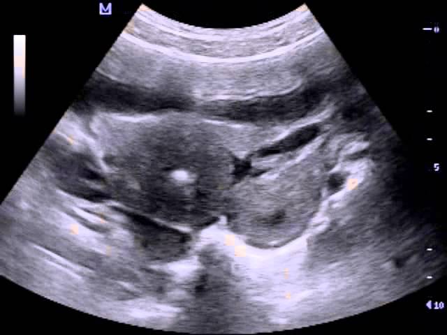
Ultrasound of the uterus, ovaries and cervix is \u200b\u200bthe most affordable way diagnosing a variety of diseases of the female reproductive system. The gynecologist sends women to this examination if there are constant and pulling pains in the lower abdomen, lumbosacral region, perineum, if menstruation is inconsistent and with other alarming symptoms.
If a woman has anxiety symptoms, she is sent for research. With the normal functioning of the pelvic organs, all indicators should be normal. During an ultrasound scan, the uzist necessarily considers:
- How is the uterus located in the pelvic area. Normally, it should be slightly tilted forward.
- What are the outlines of the uterus. Normally, they should be even and clearly visible.
- What are the sizes of the uterus.
Parameters of the uterus at reproductive age
| Group | Neck length (cm) | Neck thickness (cm) | Neck width (cm) | Uterine body length (cm) | Uterine body thickness (cm) | Uterine body width (cm) |
| There were no pregnancies | 2,9+-0,5 | 2,6+-0,4 | 2,9+-0,5 | 4,4+-0,6 | 3,2+-0,5 | 4,3+-0,6 |
| Only abortion | 3,1+-0,5 | 2,7+-0,4 | 3,1+-0,5 | 4,9+-0,6 | 3,7+-0,5 | 4,6+-0,5 |
| Childbirth 1 | 3,4+-0,6 | 2,8+-0,4 | 3,3+-0,5 | 5,1+-0,6 | 3,9+-0,5 | 5,0+-0,5 |
| Childbirth\u003e 1 | 3,7+-0,6 | 3,0+-0,5 | 3,4+-0,5 | 5,6+-0,9 | 4,3+-0,6 | 5,5+-0,5 |
Postmenopausal uterus parameters
- What is the echogenicity of the walls of the uterus. Normally, it should be uniform.
- What is the thickness and structure of the endometrium. These indicators are directly proportional to the phase of the menstrual cycle.
- What is the structure of the uterine cavity. It is considered normal if it has the same structure with even smooth edges.
When any inflammatory and pathological processes occur, there are obvious deviations from the norm of some indicator in women.
What does the small size of the uterus in women say?
 The small size of the uterus (hypoplasia) is considered a developmental disorder of the pelvic organ. Because of this, deviations in the functioning of the uterus occur. Hypoplasia can lead to infertility and the inability to bear the fetus. To confirm this diagnosis, the gynecologist sends the patient for examination. The dimensions of the ovaries, cervix, body, the thickness of the walls of the small uterus do not correspond to the established norms.
The symptoms of such a pathological deviation are:
- Amenorrhea.
- Pain during menstruation.
- Dysmenorrhea.
- Reduced attraction to the opposite sex.
- Infertility.
- Not carrying a fetus.
- Lagging in physiological development.
The normal size of the uterus in women: body length - 70-80 mm, body width - 40-50 mm, body thickness - 20-30 mm. Be sure to take into account when making a diagnosis and gynecological examination and the corresponding symptoms.
This deviation in women is diagnosed by ultrasound. Necessarily paid great attention the size of this female organ... Tests for sex hormones are also prescribed.
What does an enlarged uterus mean?
 The most common cause of an enlarged uterus is pregnancy. It should also be borne in mind that with age, the size of the uterus also becomes larger. If the process of change is observed within acceptable limits, then this is normal, but in most cases it is a sign of a pathological condition.
Signs of an enlarged uterus include:
- Constant, aching pain.
- Fluid incontinence appears.
- Discomfort during intercourse.
- Painful sensations during menstruation, as well as the presence of large blood clots.
- Bloating in the lower abdomen.
- Feeling of fullness in the abdomen.
- Back pain.
- Migraine.
- Bleeding between periods.
- Weight failure.
- Changes in the mammary glands.
- A sharp decrease in hemoglobin.
Normal sizes of the cervix
Ultrasound is prescribed to determine pathologies in the cervix. If deviations are observed, then it is imperative to urgently start treatment. The normal body length of the cervix is \u200b\u200b3.5-4 cm, and the anterior and posterior size of the cervix is \u200b\u200b2.5-3 cm. The echo structure of the cervix is \u200b\u200bhomogeneous. The endocervix of the cervix has dimensions up to 0.2-0.3 cm and is filled with mucous formations. If the ultrasound doctor detects an increase in the cervix, and the thickness also does not meet the recognized standards, then this may indicate inflammation, diseases, tumors and oncology.
Normal size of the ovaries on ultrasound
 If a woman has constant pain in the lower abdomen on the right or left side, discomfort during intercourse, ovulation is painful, then this is a reason to contact a gynecologist for a referral for an ultrasound of the ovaries. The doctor preliminarily conducts an examination.
The parameters of the ovaries should be in the following norm: width - 2.5 cm, length - 3 cm, thickness - 1.5 cm. The volume of one ovary is 2 to 8 cc. If the size, volume, thickness of the ovaries is higher than normal, then this may indicate a cyst, oophoritis. With normal performance, functioning and the absence of ovarian pathologies, the outlines should be clear and with small tubercles. The echo structure is homogeneous. In healthy ovaries, follicles of about 0.4-0.6 cm can be identified and the dominant follicle is 2.5 cm. If the thickness of the dominant is greater than the norm, this is an ovarian cyst. It is recognized by ultrasound as the formation of a liquid that is more than 2.5 cm thick.
Ovarian size at reproductive age
Ovarian size in postmenopausal women
| Neck length (cm) | Thickness (mm) | Width (mm) | Volume (cm³) | |
| Postmenopause | 25+-9 | 12+-5 | 15+-6 | 4,5+-0,9 |
| Postmenopause | 23+-9 | 11+-4 | 14+-4 | 3,5+-0,8 |
| Postmenopause | 22+-7 | 10+-4 | 13+-5 | 2,5+-0,8 |
| Postmenopause | 10+-6 | 9+-3 | 12+-4 | 1,5+-0,7 |
At first warning signs arising in the lower abdomen, it is necessary to urgently seek the advice of a specialist and undergo an ultrasound examination of the uterus, cervix, and ovaries.
Various lesions of the cervix can cause infertility either due to obstruction of the channel for sperm cells, or as a result of their death from the influence of inflammatory exudate and toxins formed in the cervix.
Inflammation of the cervix (cervicitis)(Cervicitis). Depending on the primary lesion, there are: 1) endocervicitis (endocervicitis) - inflammation of the mucous membrane of the cervix; 2) myocervicitis (myocervicitis) - inflammation of its muscle layers; 3) pericervicitis (pericervicitis) - damage to the serous cover of the cervix.
In practice, mixed forms of cervicitis are almost always found. Cervicitis, like vaginitis, can result from trauma during childbirth or after intercourse as a result of the introduction of infectious agents and invasions. Often cervicitis is a complication of colpitis or metritis and proceeds simultaneously with them.
Clinical signs.With cervicitis, flabbiness, doughiness (edema), stickiness of the mucous membrane are revealed; sometimes even light touching it is accompanied by bleeding. The cervical canal usually opens slightly and allows 1-2 fingers to pass through. Examination with a vaginal speculum can establish hemorrhages, focal or diffuse hyperemia, bleeding, accumulation of pus and mucus with flakes. If the process takes on a chronic course, hypertrophy of the folds of the mucous membrane often occurs both in the canal itself and in the vaginal part of the cervix. Cysts sometimes form in the cervical canal.
In older animals, the hypertrophied vaginal part of the cervix may take on the appearance of a cauliflower. Polypoidly overgrown folds of the mucous membrane can serve as a mechanical obstacle to the penetration of sperm into the uterine cavity; in addition, they are adversely affected by mucus, which lingers and decomposes between the folds.
Diagnosisset on the basis of vaginal and rectal examination by hand and examination of the cervix using a vaginal speculum.
Treatment.There is no specific treatment. Douching is carried out with a solution of furacilin 1: 5000, followed by lubrication of the neck with an emulsion of antibiotics, sulfonamides, suppositories or tampons are used. In acute cervicitis, accompanied by the formation of erosions, ulcers and edema, it is useful to lubricate the vaginal part of the cervix with a 5% solution of iodine, iodine-glycerin aa, synthomycin ointment, injected into the canal of the neck of the candle. After elimination inflammatory process in the uterus or vagina, inflammation stops and in the cervix. If, due to trauma or ulcerative process, the cervical canal overgrows, the animal is excluded from the uterine composition.
In case of hypertrophy of the folds for successful fertilization, it is recommended to douche the vagina with a physiological or saline-soda solution before coitus (to remove mucus and neutralize secretions) or use artificial insemination. Individual polyposis growths sitting on legs can be cut off with scissors, pulling the neck to the vulva. Parenchymal postoperative bleeding quickly stops after lubricating the wound surface with 5% iodine solution.
Induction of the cervix(Induratio cervicis) can be a consequence of chronic cervicitis, when the muscles of the cervix are replaced by connective tissue that undergoes hyalinization and even calcification. Often, cervical induration occurs after ruptures or bruises of its tissues, interstitial hemorrhages. Induction, as a rule, entails narrowing or complete obstruction of the cervical canal.
With vaginal and especially with rectal examination, the cervix is \u200b\u200bpalpated as an unevenly enlarged, compacted or stony formation. Stony hillocks alternate with compacted, resilient areas; sometimes, on the contrary, the seals are located in the grooves between the eminences (cicatricial contractions). Induction and calcification of the cervix is \u200b\u200ba direct indication for the discarding of queens. If induration is established during labor, an operative dilatation of the cervix or a caesarean section can be performed.
, Neoplasms in the cervix.Tumors in the form of sarcomas, carcinomas, fibroids and others, if they do not always create a mechanical obstacle to fertilization, then, as a rule, complicate the labor act. Tumors are localized mainly in the vaginal part of the cervix. Here, cysts and fibromas from hypertrophied folds of the mucous membrane develop especially often.
Diagnosisestablished by vaginal and rectal examination.
Treatment.Tumors developing on the vaginal part of the cervix and sitting on the legs can be removed surgically. When malignant tumors and extensive damage to the neck, the animal is discarded. Surgical intervention on the cervix is \u200b\u200brecommended only in cases where the tumor does not capture the muscle layer, remains mobile and associated only with the mucous membrane.
Preparation for surgery is normal; anesthesia - regional or local. It is advisable to dissect the perineum. The tumor is grasped with a forceps or hooked forceps and pulled out of the vagina along with the cervix. When operating under local anesthesia, an anesthetic solution is injected into the base of the tumor. An incision is made in the mucous membrane around the tumor stem with a scalpel. The edges of the incision are fixed with tweezers, and the tumor is gradually exfoliated to healthy tissues. So that after the separation of the tumor, the cervix is \u200b\u200bnot prematurely retracted into the depths birth canal, it is held with forceps. After removal of the tumor, the wound of the mucous membrane is fastened with several knotty sutures (preferably from catgut). Before suturing, it is necessary to carefully stop the bleeding by injecting or unscrewing the bleeding vessels. Sometimes, with postoperative bleeding, in order to avoid the formation of extensive hematomas, the remainder of the leg is squeezed with hemostatic forceps and left for 12-24 hours. For persistent postoperative parenchymal bleeding, a tight vaginal tamponade and vestibule are used for a period of 4-5 hours.
Small mucous polyps are successfully unscrewed with Muse forceps or cut off with scissors after preliminary stitching and tugging of the leg with a ligature.
Wrong position of the cervix.The cervix usually takes a longitudinal position, and its mouth lies in the center or deviates slightly up, down or to the sides. The position of the neck can vary depending on the physiological state of the genitals, on the tone of the abdominal muscles, and especially on the degree of filling gastrointestinal tract... Therefore, about curvature and wrong position the cervix as the cause of infertility can be talked about only when topographic deviations are strongly expressed (the cervix occupies a transverse or vertical position) and is associated with a former inflammatory process that caused the formation of adhesions of the perimetry with surrounding organs and tissues.
If, during palpation, the neck does not move due to adhesions, the animal is discarded. Kinks of the cervical canal can occur with cysts, abscesses, or trauma-related scars. They should be considered not as an independent disease, but as a sign of the disease.
Diseases of the uterus.Diseases of the uterus not only negatively affect fertility, but reduce all types of animal productivity. Inflammatory processes create unfavorable conditions in the uterus for the experience of sperm cells (spermiolysins, spermiotoxins, bacteriotoxins and bacteriolysins appear, active forms phagocytes, etc.); in addition, even after fertilization of the egg, the embryo that has fallen into the cavity of such a uterus dies. The development of inflammatory processes in the uterus during pregnancy, as well as deep morphological changes in its mucous membrane (atrophy, scars, degeneration) can lead to a violation of the connection between the fetal part of the placenta and the mother, and microbes penetrate through the damaged placental barrier into the tissues and organs of the fetus and their toxins. Sometimes pregnancy is terminated because the uterus, tightened with scars or sealed by adhesions to surrounding organs, cannot serve as a normal fetus.
The main etiological factor of uterine diseases should be considered an infection, the causative agents of which penetrate into the uterus during estrus, insemination and postpartum period... Often, uterine lesions are a complication of colpitis, cervicitis. Sometimes the causative agent of the infection enters it through the hematogenous route. Often, inflammation of the uterus is only a sign of a general disease of the body (tuberculosis, brucellosis). Depending on the virulence of microbes and the resistance of the genital tissue, signs of uterine disease can vary from obvious to subtle pathological changes by clinical examination. Therefore, the diagnosis of some lesions of the uterus is very difficult.
As a cause of infertility, there can be pathological changes in the uterus of a non-inflammatory nature in the form of endometrial hyperplasia. This hyperplasia is clinically manifested by the thickening and laxity of the uterine wall. Changes in the uterus are preceded by the formation of ovarian cysts (A.I. Ilyina).
Chronid endometritis (Endometritis chronica)- Chronic inflammation of the uterine mucosa, accompanied by more or less significant changes in the endometrium and increased activity of healthy or degenerated uterine glands. By the nature of the pathological process (clinically - by the composition of the secreted mucus), there are:
a) Endometritis catarrhalis chronica, characterized mainly by the release of serous exudate;
b) Endometritis catarrhalis et purulenta chronica, in which the discharge from the genitals is mucopurulent.
Chronic catarrhal endometritisdevelops as a consequence of the pathogenic action of the infectious agent that penetrates into the uterine cavity in the postpartum period, during coitus, artificial insemination or during the spread of the inflammatory process from the vagina and cervix.
Chronic uterine catarrh is characterized by profound changes in its mucous membrane in the form of thickening, loosening, erosion and ulceration. Connective tissue grows in the thickness of the mucous membrane; the uterine glands atrophy, and in some places they form cysts ranging in size from a pinhead to chicken eggs; individual glands or groups of them undergo hyperplasia. Along with atrophy and thinning of the mucous membrane, thickening of the folds is observed, and various shapes growths of granulation tissue turn into scars.
Typical signs: infertility, discharge from the uterus of cloudy flocculent mucus, sometimes mixed with blood. The frequency and intensity of the sexual cycles are disrupted or completely disappear. Sometimes they proceed regularly, but infertility is noted, despite numerous inseminations, or latent abortions occur in the initial stages, after which signs of the stage of arousal of the sexual cycle are revealed in the animal after 1-2 months. In the vagina, they find streaky hyperemia, an accumulation of turbid mucus of a slightly acidic reaction (pH 6-7). Usually, exudate is secreted while the animal is lying, mainly during estrus. In the equilibration stage, there is an increase and hyperemia of the vaginal part of the cervix or its displacement to the sides, up; the channel is slightly open and allows 1-2 fingers to pass.
In some cases, sexual cycles stop, there is no discharge, catarrhal exudate accumulates in the uterus.
Swabs taken from the uterus or cervical canal consist of mucus, a large number disintegrating leukocytes and ciliated epithelial cells. Rectal examination establish an uneven thickening of the walls of the horns and the body of the uterus. In places the tissue is compacted, in places flabby or fluctuates (cysts). Often the total volume of the uterus is increased; horns and body descend into abdominal cavity, and the ovaries - sometimes to the level of the pelvic floor (in mares). The motor function of the uterus is impaired: rigidity is poorly expressed or absent.
Forecastin chronic catarrhal endometritis, it is doubtful, since a number of deep morphological changes in the uterus remain irreversible.
Chronic endometritis- a kind of catarrhal, characterized by the absence of morphological changes (in a clinical study). The onset of the disease is facilitated by the use of sperm contaminated with opportunistic or pathogenic microorganisms, incorrect choice the time of insemination, at which infertility occurs and the likelihood of infection of the genitals increases.
The disease is recognized by the failure of repeated inseminations. The rhythm of the sexual cycles is usually not disturbed. During estrus, there is a profuse discharge from the genital fissure of cloudy mucus mixed with flakes. To clarify the diagnosis, mucus can be examined (see "Gynecological examination").
When saline is injected into the uterine cavity (through a reverse flow catheter), the fluid poured back from the uterus contains flakes. In the absence of clinical signs, it is advisable to perform an endometrial biopsy for histological examination. With latent endometritis, dystrophy and desquamation of the superficial epithelium, diffuse or focal accumulation of lymphoid cells, destruction of the uterine glands, severe edema of the endometrial stroma, blood filling of blood vessels are noted (A. Yu. Skripitsyn). When protracted course atrophic processes occur.
With latent endometritis, toxic substances accumulating in the uterus have a detrimental effect on sperm cells. Therefore, to restore the fertility of the animal, it is necessary to free the uterus from exudate and increase its tone. A high therapeutic effect is provided by the use of tissue therapy according to the Filatov method in combination with massage of the uterus. The drug is prepared from the liver. Sometimes positive results are obtained after washing the uterus with physiological or saline-soda solution for 1-2 hours or directly. insemination.
Ironic purulent-catarrhal endometritis -develops from acute or is caused by the activity of pyogenic microorganisms in chronic endometrial catarrh. Pathological changes are expressed quite sharply. Along with edema, severe hyperemia and hemorrhages in the uterine mucosa, more or less extensive foci of softening, purulent infiltration, degeneration, necrosis with tissue rejection appear. Often, ulceration extends to the muscle layers. The mucous membrane atrophies; its folds are smoothed out. In some places, connective tissue grows in the form of warty, mushroom-like eminences. Sometimes significant areas of the endometrium turn into a solid granulating surface or into a zone of cicatricial cords that form large folds and adhesions on the inner surface of the uterus. Cysts of various sizes are formed from the uterine glands. The uterine cavity contains mucopurulent, cloudy, liquid or thick, creamy, yellowish-white or yellow with a reddish tinge exudate.
Purulent catarrhal inflammation of the uterus is often accompanied by a deterioration in the general condition, weakening of appetite and emaciation of the animal. Fever is common. Sexual cycles drop out or become irregular (anaphrodisia and nymphomania). A white, creamy, purulent-catarrhal exudate (leucorrhoea) is periodically released from the vagina, especially plentiful during estrus.
The mucous membrane of the vagina and the orifice of the cervix is \u200b\u200bedematous, strongly hyperemic, ulcerated in places. The cervical canal is slightly open and allows 1-2 fingers to pass. During rectal examination, fluctuation (cysts), swelling, laxity of uterine tissues and cicatricial seals are established; its rigidity is expressed, weak or absent. Sometimes only certain parts of the uterus are edematous.
Persistent treatment manages to stop the development of the pathological process. However, after the disease, such profound changes in the endometrium sometimes remain that during fertilization an abortion occurs at various stages of pregnancy.
Hydrometer(Hydrometra) - a peculiar flowing catarrhal endometritis. With severe swelling of the cervix, its overgrowth or adhesions of the vagina, sometimes due to sluggishness of the muscles during twisting of the uterus, the catarrhal exudate formed in its cavity does not find an outlet and accumulates in an amount of 15-20 liters in large animals, 6-15 liters in sheep, goats and pigs and several liters in dogs. The uterus is a fluid-distended sac (Hydrometra). Accumulations in the uterus may also consist of pus (Pyometra). The wall of the uterus becomes thinner or thickens in places due to the proliferating connective tissue.
Diagnosing pyometra and hydrometer is not difficult. Sexual cycles are disrupted; the animal is often considered pregnant. Exudate is periodically or constantly released from the genitals, accumulating in the lower corner of the vulva, staining the hair on the tail and hind legs. When the cervical canal is completely closed or the vagina becomes infected, there may be no discharge.
Vaginal examination reveals hyperemia, edema and ulceration of the mucous membrane of the vagina and cervix, cicatricial contractions or adhesions. During rectal examination, a tightly fluctuating or somewhat dough uterus is probed. Its body and horns are evenly stretched by the contents; the wall is thinned or, conversely, in some places has a skin-like consistency. The uterus descends into the abdominal cavity; shift - down and the ovaries. With a large increase in the uterus, vibration of the middle uterine arteries, but it is weaker than during pregnancy, and is usually equally pronounced on both sides. Hydro - and pyometra can be mistaken for a twin pregnancy. But changes in the wall of the uterus, absence of a fetus, symmetrical enlargement of the horns, equal strength pulsation of the right and left uterine arteries, general state the animal give the basis for the correct diagnosis. In cows, with a uniform increase in the uterus, the placenta is not palpable (absent); one or both ovaries contain large yellow bodies.
Forecastpoor, the fertility of the animal is not restored.
Chronic endometritis in small animals.Inflammation of the uterus protects mainly in the form of catarrhal and purulent-catarrhal endometritis and develops when the causative agent is introduced after childbirth, during coitus, or with uterine atony. Pathological changes are the same as in large animals,
With purulent endometritis, capturing the deep layers of the uterus, the temperature rises, the general condition worsens, diarrhea appears; animals often die from cachexia. Characteristic feature - discharge from the genitals of mucous or mucopurulent exudate, yellow or brown color... Only a full uterus can be felt through the abdominal wall in sheep and goats. In a combined study by the external rectal method, a thickened dense or fluctuating uterus is installed. In dogs, cats and rabbits, the uterus can be felt through the abdominal walls and fluctuations are established, cord-like, unevenly or evenly thickened and compacted areas of the horns in the form of tuberosities located along the spine.
In order not to confuse the uterus with the intestinal loops, the bifurcation of the horns should be taken as a landmark. To facilitate the examination, a probe or finger is inserted into the vagina, from the end of which, as from the starting point, palpation begins through the abdominal walls. In large pigs, the uterus can be probed rectally. In doubtful cases, diagnostic laparotomy is appropriate. The abdominal cavity can be opened along the white line or from the side. The incision is made so that 1-2 fingers pass through it. Preparation for laparotomy is carried out taking into account the possible detection of direct indications for total amputation of the uterus. The reproductive function of the animal is not restored; both dogs and cats are often fatal. Parenteral ichthyol, oxytocin or pituitrin, tissue preparations, etc. are used as therapeutic measures. In case of pyometra and deep changes, the uterus in small animals is amputated.
Myometritis (Myometritis) -inflammation of the muscular membrane of the uterus, resulting from severe endometritis, less often - the penetration of the pathogen by the hematogenous route. Intermuscular connective tissue grows, replaces muscle fibers and undergoes hyaline or amyloid degeneration. In places in the wall of the uterus, lime salts are deposited, abscesses develop, opening into the uterine cavity or encapsulating. For myometritis, a violation of the motor function of the uterus is characteristic.
On rectal examination, the uterus feels thickened, sometimes unevenly bumpy, in places of a stony consistency. Her body and horns descend into the abdominal cavity. These phenomena are often associated with signs of endo - and perimetritis.
Treatmentchronic myometritis does not give positive results; animals are culled from the brood herd, as they remain sterile or undergo routine abortions.
Rebirth of the uterus.Degenerative processes in the uterus in the form of amyloid infiltration, hyaline degeneration of interstitial and connective tissue overgrown with myometritis is a very rare phenomenon. More often there is a thickening of the uterine wall due to the development of fibrous connective tissue in its muscle layers as a result of chronic myometritis.
With rectal examination, an increase in the uterus is established; its surface is uneven, bumpy, dense, in places of a stony consistency. The rigidity is impaired. The ovaries are reduced in volume, do not contain yellow bodies and follicles (atrophy). As a rule, the degeneration of the uterus is accompanied by anaphrodisia.
Forecastadverse. Animals are culled.
Uterine atrophyobserved in old animals with prolonged hydro- and pyometers, after staying in the uterus of a macerated or mummified fetus, with lesions of the ovaries (cysts, sclerosis).
In the uterus, the number and size of muscle fibers decrease, which are partially replaced by connective tissue. Sexual cycles stop.
The diagnosis is made on the basis of the results of rectal examination, as well as taking into account the history and age of the animal. The uterus and its neck do not correspond to the size of the animal. The uterine horns are small, thin, flabby or dense, sometimes they are enlarged due to the accumulation of mucus in their cavity or the formation of connective tissue growths in the endometrium. Stiffness is absent and does not appear even after vigorous massage of the uterus.
The ovaries are reduced, dense, do not contain corpus luteum and follicles. When diagnosing, it is necessary to exclude alimentary, climatic and operational infertility.
Forecastadverse; fertility is usually not restored. As a treatment, you can experience hormone stimulation, massage of the uterus and ovaries.
Neoplasms in the uterusanimals are rare; they are found mainly in cows and dogs. By histogenesis and pathological structure, tumors in the uterus can be very diverse: sarcomas, carcinomas, fibroids, leiomyomas, fibroleiomyomas, fibrosarcomas, etc.
Diagnosisset by palpation, and in small animals - after diagnostic laparotomy.
It is possible to remove the affected tumor of the uterine horn or part of it only in small animals (dogs, cats). Large animals are culled for meat upon diagnosis.
Inflammation of the fallopian tube.The condition of the oviduct is of great importance for the fertility of the animal. Many large and small folds of the mucous membrane protrude into the lumen of the oviduct; therefore, the development of an inflammatory process in the fallopian tube, accompanied by swelling of the mucous membrane and adhesions of its folds, leads to the formation of closed cavities, which contain exudate. Under the influence of the toxins present in the latter, sperm, egg and zygote die.
Inflammation of the oviduct and its consequences in the form of degeneration of individual layers of the tube interfere with the advancement of the egg and often serve as an insurmountable obstacle on the way of sperm to the egg cell. Insignificant in size, elusive in a clinical study, lesions of the tubes can cause infertility of the animal in normal condition and the correct functioning of all other parts of the reproductive apparatus.
Lesions of the oviduct can be limited to the mucous membrane (Endosalpingitis), capture the muscle layer (Myosalpingitis) or the serous membrane of the tube (Perysalpingitis). In fact, there are no methods of thorough clinical research that would allow to identify lesions of individual layers of the oviduct. Clinically, only processes accompanied by morphological changes in the tube in the form of accumulations of exudate or connective tissue cicatricial growths are detected. Therefore, we will limit ourselves to considering only the main lesions of the tubes.
Acute and chronic catarrhal salpingitis(Salpingitis catarrhalis acu-ta et chronica). Endosalpingitis - inflammation of the mucous membrane of the tube develops as a complication of endometritis, peritonitis due to trauma or metastases. The essence of the process lies in swelling, the appearance of small hemorrhages and leukocyte infiltration of the mucous membrane. Its epithelium degenerates and sloughs off in places. Edema of the folds of the mucous membrane leads to the formation of closed cavities, more or less strongly stretched by secretion, or to complete obstruction of the tube with filling of its lumen with serous-catarrhal discharge.
In the chronic course of the disease, the epithelium of the tube degenerates, its folds thicken and thicken due to the proliferation of connective tissue. The tops of the folds, devoid of integumentary epithelium, grow together, and closed cavities remain in the intervals between them. Sometimes cicatricial contractions cause strictures and even complete clogging of the tubes in one or more places. The closed sections of the tubes are stretched by the inflammatory exudate into cystic swellings. As the pathological process progresses, the degenerated mucous membrane begins to secrete a mucous-serous fluid that overflows the tubes. The process ends with dropsy of the tubes (Hydrosalpinx).
With catarrhal-hemorrhagic inflammation or after trauma (with a rough rectal examination), a significant amount of blood (haemosalpinx) can accumulate in the lumen of the tube.
Acute and chronic purulent salpingitis(salpingitis purulenta acuta et chronica). Purulent salpingitis usually arises from catarrhal and often proceeds in the form of purulent-catarrhal inflammation. Purulent salpingitis is characterized by profound changes in the mucous membrane. Erosions and ulcers appear on its surface, sometimes purulent-fibrinous and diphtheritic deposits. The pathological process captures the muscle layers. In a chronic course, scar tissue develops and the lumen of the oviduct is obliterated. Purulent exudate accumulates in the pipe canal white creamy consistency or liquid greenish or yellowish serous-catarrhal masses. The contents can stretch the pipe, merging into one continuous fluctuating bladder or into several abscesses along the pipe.
Normally, the oviducts in mares and cows are not palpable during rectal examinations. Therefore, the diagnosis of mild forms of endosalpingitis is impossible. With the exclusion of lesions of other parts of the genital apparatus, it is necessary to make a presumptive diagnosis of obstruction of the tubes.
Attempts by some clinicians to identify the patency of the tubes in cows and mares by blowing them did not give valuable diagnostic results.
With hydro- and pyosalpingitis, a round or oval fluctuating bladder is placed by feeling between the ovary and the apex of the uterine horn. In the presence of purulent contents in the tube, the animal reacts strongly to palpation. Sometimes, along the pipe, several cyst-like fluctuating swellings ranging in size from a pea to a pigeon's egg and more are felt.
Endomyoperisalpingitis(Salpingitis nodosa) - chronic inflammation oviduct, accompanied by the proliferation of connective tissue replacing muscle tissue and forming dense cords and nodes along the tube (induration). With such a strong lesion, the patency of the oviducts is disrupted and they often adhere to the ovary and surrounding tissues. Chronic salpingitis occurs as a complication of chronic catarrhal or purulent salpingitis, and in cows - after pressing the corpus luteum, as well as when the oviduct is damaged by tuberculosis.
By palpation through the rectum in the ligaments between the ovary and the uterine horn, a dense, even stony consistency, tuberous cord is easily felt. Sometimes adhesions and connective tissue growths surrounding the ovary are detected. In small animals, diagnosis is only possible through laparotomy.
Treatmentsalpingitis is inappropriate. An animal with unilateral salpingitis is considered conditionally fit for reproduction; with bilateral damage to the tubes, females are discarded. In case of dropsy of the tube, you can experience treatment with its massage, but it is very difficult to count on the restoration of patency.
Diseases and disorders of the ovarian function.The anatomical structure of the female's body, the activity of all its organs and tissues are in close functional connection with the ovary. In turn, the general condition of the body, the work of its organs, especially endocrine and nervous systems, are reflected in the morphology and function of the ovaries. Therefore, it is often difficult to establish a specific cause of infertility: it has to be found not only outside the reproductive apparatus, but also outside the body, during external environment, extremely strongly affecting the state of the ovaries.
In a number of cases, morphological changes and disorders of the ovarian function, disrupting the activity of the reproductive apparatus as a whole and caught in a clinical study, are themselves only a consequence of distant common reasons, a sign of a general disease of the body or the result of abnormal conditions of its existence. On this basis, we divide all lesions of the ovaries into two groups: 1) diseases of the ovary, developing as a result of the penetration of the causative agent of infection, trauma; 2) functional disorders of the ovary, which are a sign of damage to other organs and systems of the body or the result of errors in the care of the female, her maintenance, feeding and exploitation.
Ovariite (oophoritis) (ovaritis, s. oophoritis) develops with the introduction of the th causative agent of infection with inflammation of the uterus, tubes, peritoneum and other organs; after squeezing the corpus luteum, cysts, massage of the ovary and other traumatic influences. Less commonly, oophoritis occurs due to the penetration of the pathogen by the hematogenous route.
Oophoritis can manifest itself in the form of serous, hemorrhagic, purulent inflammation in acute or chronic course. However, in a clinical study, the nature of the inflammatory process usually cannot be established.
NP Omelchak, using endoscopy and rectal examination in cows, found that the first signs of inflammation in the ovary appear after 8-72 hours, less often 6 days after exposure to the cause. So, after a rough massage of the ovaries, signs of inflammation appear after 10-15 hours, after enucleation of the corpus luteum and crushing of cysts - after 12-18 hours, after the introduction of microbes into the ovarian tissue - after 54-72 hours and with endometritis - by 6-14 th day. More often, the disease begins with serous inflammation, which after 2-5 days turns into purulent or hemorrhagic, the latter can develop independently. On palpation of the affected ovary, the animal shows anxiety. Abscesses in the ovary appear 5-7 days after the onset of the disease. The decrease in the inflammatory response occurs on the 7-10th day.
Treatment.Apply novocaine therapy, antibiotics; it is useful to use heat (hot douching), ozokeritotherapy and mud therapy (vaginal tampons, applications on lumbar region). E.V. Ilyinsky recommends intramuscular injection of penicillin with streptomycin at 1500-2000 IU or tetracycline with monomycin, respectively, 1500 and 2000 IU per 1 kg of animal weight (2-4 times a day for 3-5 days).
Purulent oophoritis(oophoritis purulenta) is characterized by the formation of abscesses in the ovary tissues ranging in size from a pea to a chicken egg and more. Ulcers can develop as a complication of purulent processes in the uterus, tubes, or as a result of metastases. They are often opened into the abdominal cavity or encapsulated.
On palpation, the ovary appears enlarged; fluctuation is sometimes felt; when feeling the ovary, the animal is very worried (which does not happen when feeling the cysts). The corpus luteum and follicles are not detected. Purulent ovariitis, as a rule, is accompanied by a vivid general reaction of the body in the form of oppression, refusal to feed. Along with changes in the ovary, it is almost always possible to establish lesions of the uterus, tubes or other organs adjacent to the ovary. The rhythm of the sexual cycles is disrupted in the form of anaphrodysia.
Treatmentsymptomatic.
Chronic parenchymal ophoritis(oophoritis parenchymatitosa chronica) develops from an acute form of parenchymal or purulent oophoritis and is a deep tissue change - the replacement of ovarian elements with connective tissue, which undergoes hyalinization and calcification. The organ increases, it becomes, as it were, stony, and its surface is bumpy due to the appearance of powerful scars. With unilateral damage to the ovaries, the rhythm of the sexual cycles may not be disturbed; with bilateral oophoritis, anaphrodisia is observed. By palpation, an increase in the ovary is established, its dense, in places cartilaginous consistency. Sometimes cicatricial contractions are felt. No signs of follicles and yellow bodies are found. Often the enlarged ovary descends.
To give an accurate conclusion about the nature of the disease, it is necessary to examine the animal 2-3 times at intervals of 25-30 days.
Chronic parenchymal oophoritis causes irreversible changes. With bilateral damage to the ovaries, the animal is discarded.
Chronic interstitial oophoritis,ovarian sclerosis (oophoritis interstitalis.ehronica). With interstitial inflammation, connective tissue grows in the ovary and is hyalinized. The ovarian parenchyma atrophies; the tunica albuginea is strongly thickened. The organ decreases, its surface becomes uneven, bumpy. Bilateral interstitial inflammation of the ovaries is especially common in older cows. Sclerosis can sometimes only be in part of one ovary.
In animals, anaphrodisia is noted. Palpation reveals dense ovaries with an uneven surface; reduced in cows to an acorn or bean. Often, ovarian sclerosis is accompanied by uterine atrophy. Females with partial or unilateral ovarian sclerosis are considered fertile. Bilateral damage to the ovaries causes infertility, so females with this disease are discarded from the breeding stock.
Ovarian atrophy.Bilateral atrophy of the ovaries in combination with atrophy of the uterus often occurs in old, malnourished, but high milk yield cows. Ovarian atrophy is recognized by the absence of sexual cycles in animals. In rectal examination, the ovaries have an elastic-elastic consistency and size: in cows - with a bean and even a pea, in mares - with a pigeon's egg.
Ovarian atrophy is accompanied by deep and irreversible tissue changes (absence of follicles, decrease in interstitial tissue, desolation and hyalinization of blood vessels). Animals are culled from the breeding stock. Gonadostimulating agents, ovarian and uterine massage, electrophysiotherapy, and other treatments can be tested to treat valuable breeding females, albeit with little hope of a positive outcome.
It should be borne in mind that alimentary, operational, climatic and other forms of infertility can stimulate ovarian atrophy. Therefore, one can speak of ovarian atrophy only after excluding a symptomatic decrease in ovarian function in animals.